

Consider the papercut—a minor injury best known for the disproportionate amount of pain it can cause. That a wound so inconsequential can sting so terribly is curious, but perhaps even more surprising is the fact that it heals at all.
To heal a wound, even one as trivial as a papercut, the cells involved in the repair must migrate to precisely where they’re needed at the site of injury. Determining where to go requires interpretation of a substantial number of factors, including chemical signals, fluid dynamics, mechanical properties, and more. Cells also migrate in other, similarly complex circumstances, from embryonic development to cancer metastasis.
Most of what we know about how these feats are controlled has come from in vitro studies with cells growing in essentially two-dimensional layers in Petri dishes. Although this research continues to provide invaluable insights, in vitro studies alone can’t give a complete understanding because cells grown in such conditions don’t move exactly the way cells in the body do. In GENETICS, researchers David Sherwood and Julie Plastino review how studies using the microscopic nematode Caenorhabditis elegans are improving our understanding of cell migration.
Sherwood and Plastino explain several reasons that C. elegans is an especially good model for this kind of research. The worms’ simplicity makes them easier to study, and many aspects of cell migration are conserved from C. elegans to humans. C. elegans’ status as one of the best-established model organisms means researchers can take advantage of established protocols to genetically manipulate and study the worms. The worms are transparent, making tracking cell migration less complicated. Also helpful is the fact that the worms develop in a very predictable, choreographed way: hermaphrodites (which, in worms, are essentially females) have precisely 959 body cells, while males have 1031 body cells.
Cell migration in the worms takes many forms, some of which the authors discuss in the review. In one example, they describe how cells penetrate basement membranes, which are thin but tough layers of extracellular matrix found in animals. In humans, basement membranes attach surface cells—such as the top layers of skin and the lining of blood vessels—to the underlying connective tissue. Basement membranes act in part as cellular gatekeepers, preventing cells from escaping tissues. Human basement membranes, however, must be punctured in a variety of circumstances, such as when new blood vessels need to grow through the membranes and when cancer cells spread.
Most C. elegans tissues are encased in basement membranes, and as in humans, these membranes must sometimes be breached. During the worms’ development, a type of uterine cell called an anchor cell burrows through a basement membrane that separates the vulva from the uterus. This infiltration links the organs together, allowing the worms to mate and lay eggs.
Studying how anchor cells invade the basement membrane in C. elegans has produced numerous insights into human cell migration—both normal and pathological. In 1989, scientists first reported in vitro observations of invasive tendrils they called invadopodia protruding from cells grown on a simplified extracellular matrix, including cells from established cancer cell lines and primary tumor cells.
Later, C. elegans researchers found invadopodia on anchor cells penetrating the basement membrane, providing the first evidence that the structures exist not only in cells grown in vitro, but also in a whole animal. They also discovered some discrepancies between the characteristics of invadopodia in cells grown in vitro and C. elegans cells, which could reflect differences in how invadopodia behave when subjected to the full range of stimuli present in vivo.
In the review, Sherwood and Plastino highlight many more discoveries about cell migration that have been made—and continue to be made—using C. elegans. All evidence indicates that C. elegans will remain a valuable asset to the field, serving not only as a link between in vitro studies and investigations of more complex animals such as humans, but also as a rich source of fundamental discoveries in itself.
Sherwood, D.; Plastino, J.
Genetics, 208(1), 53-78.
DOI: 10.1534/genetics.117.300082
http://www.genetics.org/content/208/1/53
Nicole Haloupek is a freelance science writer and a recent graduate of UC Berkeley's molecular and cell biology PhD program.
View all posts by Nicole Haloupek »Read more in
-

Thank you, GSA community!
Thank you for being a member of the Genetics Society of America! As GSA’s current president, I am writing to tell you about Society projects and initiatives that we hope you will find useful in advancing your science and your career. Scientific research is a collaborative and exciting endeavor. Scientific societies like GSA exist to…
-

Where are they now? Rosalind Franklin Young Investigator Award recipients share updates on their research
Rosalind Franklin Young Investigator Award applications are open–make sure you submit your application or nomination of a colleague by September 30, 2024.
-

University of Minnesota researchers map genome of the last living wild horse species
The study, published in G3: Genes|Genomes|Genetics, is part of larger conservation efforts to save Przewalski’s horse.
-

Congratulations to the Spring 2024 DeLill Nasser Awardees!
GSA is pleased to announce the recipients of the DeLill Nasser Award for Professional Development in Genetics for Spring 2024! Given twice a year to graduate students and postdoctoral researchers, DeLill Nasser Awards support attendance at meetings and laboratory courses. The award is named in honor of DeLill Nasser, a long-time GSA supporter and National Science Foundation…
-

Carolyn Damilola: an NFS Rising Scientist on a lifelong quest to learn more
Carolyn Damilola is an NFS Rising Scientist from Nigeria doing respiratory system research and paving the way for scientists from underrepresented communities through mentorship.
-
What does a good microgrant proposal look like?
Members of the Microgrant Review Committee share their tips for a successful proposal.
-

The first piece of the facial recognition puzzle
New research in GENETICS gives a first peek at the molecular pathway involved in recognizing faces.
-

New Senior Editor Amy MacQueen joins GENETICS
A new senior editor is joining GENETICS in the Genome Integrity and Transmission section. We’re excited to welcome Amy MacQueen to the editorial team.
-

Block party on the zebrafish sex chromosome
Research in G3 identifies a gene regulatory block of the zebrafish genome responsible for overseeing the maternal-to-zygotic-transition.
-

Unraveling the mysteries of duckweed: epigenetic insights from Spirodela polyrhiza
Research published in G3 offers insight into the impact of DNA methylation on clonal propagation in asexually reproducing plants.
-

A microbiologist’s quest to understand CRISPR in bacterial self-defense
2024 Genetics Society of America Medal recipient Luciano Marraffini determined how CRISPR-Cas systems destroy genetic targets with precision, paving the way for gene editing technology development.
-
Unlocking mysteries of trait and disease heritability in dogs
2024 Edward Novitski Prize recipient Elaine Ostrander, a pioneer of the domestic dog model, discovered numerous genes affecting dog size, morphology, behavior, and disease susceptibility—many of which have relevance in humans.
-
GSA and collaborators Personal Genetics Education & Dialogue and Reclaiming STEM Institute launch NSF-funded BIO-LEAPS project to support culture change in genetics
We are thrilled to announce that the Genetics Society of America (GSA) is collaborating with the Personal Genetics Education & Dialogue (PGED) based in the Department of Genetics at Harvard Medical School, and the Reclaiming STEM Institute (RSI) on a Leading Culture Change Through Professional Societies of Biology (BIO-LEAPS) grant from the U.S. National Science…
-

Daman Saluja: Navigating Science and Policy in India
In the Paths to Science Policy series, we talk to individuals who have a passion for science policy and are active in advocacy through their various roles and careers. The series aims to inform and guide early career scientists interested in science policy. This series is brought to you by the GSA Early Career Scientist…
-

A fly geneticist’s journey into discovering rules of organ development
2024 George W. Beadle Award recipient Deborah Andrew discovered new genes and pathways in Drosophila salivary gland organogenesis. Now, her work can help optimize cell secretion in therapeutic applications and fight malaria.
-

Małgorzata Gazda: How receiving the DeLill Nasser Award helped her land her dream job
Have you ever experienced an event that changes the course of your life, or in this case, your career? Małgorzata (Gosia) Gazda is Assistant Professor at the University of Montreal and in 2022, she received the DeLill Nasser Award for Professional Development in Genetics, which she used to attend and present at the 2022 Population,…
-

Hongyu Zhao joins GENETICS as new Senior Editor
A new senior editor is joining GENETICS in the Statistical Genetics and Genomics section. We’re excited to welcome Hongyu Zhao to the editorial team.
-

GSA Member Julio Molina Pineda Receives DeLill Nasser Award, Shines at TAGC 2024
“At any career stage, the GSA membership is an amazing investment for any genetics professional!” Julio Molina Pineda is a PhD Candidate in Cell and Molecular Biology and a Research Assistant at the University of Arkansas, and a Doctoral Academy Fellow at the Lewis Lab. In 2023, Julio was awarded the DeLill Nasser Award for…
-
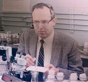
In Memoriam: Ellsworth Herman Grell (1932–2023), a pioneer of Drosophila genome engineering and annotation
Ellsworth (Ed) Grell blessed the Drosophila community through three enduring legacies: as a pioneer of chromosome mechanics, as a primary organizer and synthesizer of genetic knowledge in Drosophila, and as a graceful mentor to those fortunate to have known him personally. Ed grew up in rural Nebraska, completed his undergraduate studies at Iowa State, and…
-

Congratulations to the #Fungal24 Poster Award winners!
We are pleased to announce the recipients of the GSA Poster Awards for posters presented at the 32nd Fungal Genetics Conference! Undergraduate and graduate student members of GSA were eligible for the awards, and a hard-working team of judges made the determinations. Congratulations to all! Felicia Ebot Ojong, The University of Georgia My research is focused…
-
Poster presentation tips for TAGC 2024
You’ve been selected to present a poster at The Allied Genetics Conference 2024 in March—you’ve celebrated, made plans to attend, now what? This is an exciting opportunity to showcase your research and engage with fellow members of the genetics community, so you want to make sure you’re prepared. We wanted to offer you some tips…
-

Maximize your TAGC 2024 experience
A guide to all that National Harbor & DC have to offer Are you joining us for The Allied Genetics Conference 2024 in March? Make the most of your #TAGC24 experience in National Harbor! We know the science will keep you busy, but you deserve to unwind and have some fun, so we’ve curated a…
-
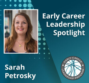
Early Career Leadership Spotlight: Sarah Petrosky
We’re taking time to get to know the members of the GSA’s Early Career Scientist Committees. Join us to learn more about our early career scientist advocates. Sarah PetroskyMultimedia SubcommitteeUniversity of Pittsburgh Research Interest I am interested in understanding adaptation that has been happening recently in populations by dissecting the ways that genes underlying an adaptation…
-
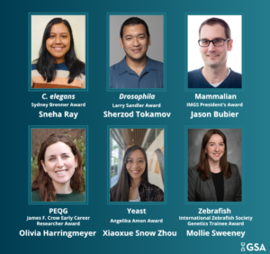
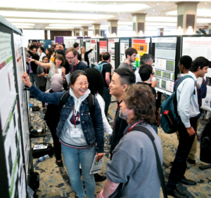
TAGC 2024 Early Career Award Winners
GSA is pleased to announce the winners of the early career awards presented at The Allied Genetics Conference 2024. These awards are specific to particular TAGC communities and recognize early career scientists’ outstanding work on their respective research organisms. The awardees will present their talks in keynote sessions at TAGC 2024. Don’t miss the opportunity…
-

Preeminent geneticists recognized with revamped GSA Awards
In 2022, GSA’s Board of Directors launched an audit to review the five major awards conferred by the Society. Today, we are thrilled to announce the recipients of the reimagined GSA Awards, including the new Genetics Society of America Early Career Medal. The scientists honored this year are recognized by their peers for their outstanding…
-
Fly Board funds outreach programs to spread the word about Drosophila research
In 2020, the Fly Board voted to use part of its reserve fund to support efforts to increase trainee participation as well as equity and diversity in the Drosophila community. An awards committee decides how the money will be spent each year, and from 2020–2022, the committee posted a very broad call for applications from…
-

New members of the GSA Board of Directors: 2024–2026
We are pleased to announce the election of four new leaders to the GSA Board of Directors: 2024 Vice President/2025 President Brenda Andrews Professor, University of Toronto It’s an honor to continue my association with the Society by serving as Vice President of the Board of Directors. I have broad knowledge of the ongoing activities…
-

Building tools and a community: How a yeast geneticist transformed medical mycology research
Aaron Mitchell, recipient of the 2026 George W. Beadle Award, built an entire career around developing and disseminating new tools to study pathogenic fungi and training the next generation of mycologists.
-

Landing a faculty position: Jiae Lee
Interviews from newly appointed faculty members shed light on the path to landing a faculty position.
-

Beyond a single reference genome: Lessons from the Human Genome Diversity Project
A new Perspectives article in GENETICS revisits the Human Genome Diversity Project through conversations with its early leaders, highlighting key lessons for today’s population geneticists.