

Mitochondria cell-autonomously regulate the secretion of neuropeptides in C. elegans.
Neurons are hard-working cells that need a lot of energy to do their jobs, so it’s no surprise that they are highly dependent on their mitochondria to function properly. Yet these organelles do much more for cells than simply produce energy. In GENETICS, Zhao et al. report on how mitochondria are directly involved in regulating the secretion of neuropeptides.
In a previous paper, the authors found that disruption of the gene ric-7 caused decreased neuropeptide secretion and locomotion defects in C. elegans—but the mechanism underlying these phenotypes was unclear because little was known about the function of ric-7. After another group demonstrated that ric-7 is required for the long-distance transportation of mitochondria from the neuron’s cell body into its axons, Zhao and colleagues hypothesized that disrupting mitochondrial transport might be the mechanism by which ric-7 defects cause neuronal phenotypes.
To test this, the authors expressed a chimeric kinesin construct, kin-Tom7, in ric-7 mutant axons. This chimera is a kinesin protein fused to a mitochondrial membrane protein. A prior study showed that kin-Tom7 restored transport of mitochondria to the axons of ric-7 mutants but did not affect other cellular functions. The authors showed that kin-Tom7 also rescues the ric-7 mutation-impaired neuropeptide secretion and locomotion defects, suggesting that improper mitochondrial transport was indeed the cause of neuronal defects in ric-7 mutants.
Because mitochondria are involved in so many cellular processes, the authors wondered which function(s) of axonal mitochondria might be necessary for neuropeptide secretion. Using selected mutant worms, they found that disrupting oxidative phosphorylation decreased neuropeptide secretion—but impairing mitochondrial calcium uptake didn’t.
Impaired oxidative phosphorylation can cause increased levels of reactive oxygen species (ROS) and hypoxia, so the authors suspected that these stress states might be involved in neuropeptide secretion. Indeed, they found that impairing the function of ROS detoxification enzymes—an alternative way to increase ROS—and growth in hypoxic conditions both led to decreased neuropeptide secretion.
Further investigation showed that the effects of axonal mitochondria on neuropeptide secretion were mediated by the hypoxia-inducible factor HIF-1, which is central to the response to hypoxia in C. elegans. Worms with constitutively active HIF-1 had lower secretion of neuropeptides, but this could be reversed by turning HIF-1 “off” again through other transgenic manipulations. Crucially, combining the constitutively active HIF-1 with the ric-7 defect in a double mutant had no additional phenotypic effects, suggesting the two proteins act in the same pathway. Consistent with this, hif-1 null mutations restored neuropeptide secretion in ric-7 mutants.
Together, these results support the idea that mitochondria regulate neuropeptide secretion in part by modulating ROS production and the hypoxic stress response. These findings could provide a mechanism by which the biochemical conditions within a neuron alter communication between neurons to trigger more widespread changes in the nervous system.
CITATION:
Tongtong Zhao, Yingsong Hao, Joshua M. Kaplan
Genetics September 2018 210: 275-285; https://doi.org/10.1534/genetics.118.301014
Science Writing and Communications Intern, Genetics Society of America.
View all posts by Marisa Wexler »Read more in
-
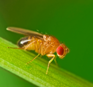
In Memoriam: Ellsworth Herman Grell (1932–2023), a pioneer of Drosophila genome engineering and annotation
Ellsworth (Ed) Grell blessed the Drosophila community through three enduring legacies: as a pioneer of chromosome mechanics, as a primary organizer and synthesizer of genetic knowledge in Drosophila, and as a graceful mentor to those fortunate to have known him personally. Ed grew up in rural Nebraska, completed his undergraduate studies at Iowa State, and…
-

Congratulations to the #Fungal24 Poster Award winners!
We are pleased to announce the recipients of the GSA Poster Awards for posters presented at the 32nd Fungal Genetics Conference! Undergraduate and graduate student members of GSA were eligible for the awards, and a hard-working team of judges made the determinations. Congratulations to all! Felicia Ebot Ojong, The University of Georgia My research is focused…
-
Poster presentation tips for TAGC 2024
You’ve been selected to present a poster at The Allied Genetics Conference 2024 in March—you’ve celebrated, made plans to attend, now what? This is an exciting opportunity to showcase your research and engage with fellow members of the genetics community, so you want to make sure you’re prepared. We wanted to offer you some tips…
-

Maximize your TAGC 2024 experience
A guide to all that National Harbor & DC have to offer Are you joining us for The Allied Genetics Conference 2024 in March? Make the most of your #TAGC24 experience in National Harbor! We know the science will keep you busy, but you deserve to unwind and have some fun, so we’ve curated a…
-
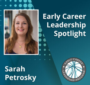
Early Career Leadership Spotlight: Sarah Petrosky
We’re taking time to get to know the members of the GSA’s Early Career Scientist Committees. Join us to learn more about our early career scientist advocates. Sarah PetroskyMultimedia SubcommitteeUniversity of Pittsburgh Research Interest I am interested in understanding adaptation that has been happening recently in populations by dissecting the ways that genes underlying an adaptation…
-
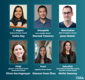
TAGC 2024 Early Career Award Winners
GSA is pleased to announce the winners of the early career awards presented at The Allied Genetics Conference 2024. These awards are specific to particular TAGC communities and recognize early career scientists’ outstanding work on their respective research organisms. The awardees will present their talks in keynote sessions at TAGC 2024. Don’t miss the opportunity…
-

Preeminent geneticists recognized with revamped GSA Awards
In 2022, GSA’s Board of Directors launched an audit to review the five major awards conferred by the Society. Today, we are thrilled to announce the recipients of the reimagined GSA Awards, including the new Genetics Society of America Early Career Medal. The scientists honored this year are recognized by their peers for their outstanding…
-
Fly Board funds outreach programs to spread the word about Drosophila research
In 2020, the Fly Board voted to use part of its reserve fund to support efforts to increase trainee participation as well as equity and diversity in the Drosophila community. An awards committee decides how the money will be spent each year, and from 2020–2022, the committee posted a very broad call for applications from…
-

New members of the GSA Board of Directors: 2024–2026
We are pleased to announce the election of four new leaders to the GSA Board of Directors: 2024 Vice President/2025 President Brenda Andrews Professor, University of Toronto It’s an honor to continue my association with the Society by serving as Vice President of the Board of Directors. I have broad knowledge of the ongoing activities…
-
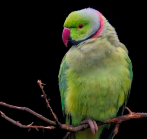
Parrot plumage study aids breeders and endangered natural populations
Yellow coloration is maladaptive in the wild but prized by breeders. People have long been fascinated with birds, which exhibit one of the widest ranges of coloration among vertebrates. Parrots, in particular, have captivated humans by their ability to mimic human speech and spectacular plumage. Brightly colored feathers are used primarily to attract mates, intimidate…
-

Early Career Leadership Spotlight: Sarah Gilmour
We’re taking time to get to know the members of the GSA’s Early Career Scientist Committees. Join us to learn more about our early career scientist advocates. Sarah GilmourMultimedia SubcommitteeStowers Institute for Medical Research Research Interest Questions of evolution have always fascinated me. I am extremely fortunate to be starting out my research career in a…