

Guest post by Beata Edyta Mierzwa. GSA-art features the creative works of scientists, particularly geneticists. Read more about the series from GSA President Stan Fields. If you would like to submit your own work or nominate someone else’s, please send an email GenesToGenomes@genetics-gsa.org with “GSA-Art” in the subject line.
I used to draw a lot—mostly portraits of my friends—before starting my studies in molecular biology at the University of Vienna. I went to Switzerland for my Master’s thesis at the ETH Zürich, and recently finished my PhD in Daniel Gerlich’s group at the Institute of Molecular Biotechnology (IMBA) in Vienna.
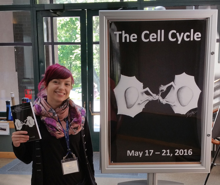
During my PhD, I realized that showing a drawing as part of my scientific presentations sparked interest and tended to stay in people’s memories. This is one of the initial reasons that I began making science-themed art. I have been very lucky to be part of an institute that encourages creativity, with yearly Art & Science contests and many other opportunities to present my art. I recently had the great honor to have my drawing selected for the cover and abstract book of the Cell Cycle Meeting in Cold Spring Harbor, and I was overwhelmed by the positive feedback I received. It was an incredibly rewarding experience that encouraged me to start creating art work for other people’s research as well as my own. You can find a complete gallery on my website (www.beatascienceart.com).
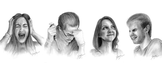
When devotion begets emotion – the life of a PhD student. These portraits of my fellow PhD students illustrate the intense emotions researchers face in everyday life in the lab. These drawings were part of a contribution to the Art & Science contest at the Vienna Biocenter, for which my team received the first prize in 2013.
Inspirations
I have always loved art, and have always loved science. I realized that combining these passions creates a unique way to communicate science.
With my illustrations, I highlight fundamental scientific aspects in an unconventional and memorable way. I want to add some creativity to the conventional forms of scientific communication, with the aim to spark interest inside and outside the scientific community. Every drawing is an experiment!
To separate our DNA during cell division, the mitotic spindle pulls the duplicated chromosomes towards opposite poles of the cell. This hand-drawn illustration shows how chromosomes align in the equator of the cell during metaphase before they are separated into each of the two daughter cells.
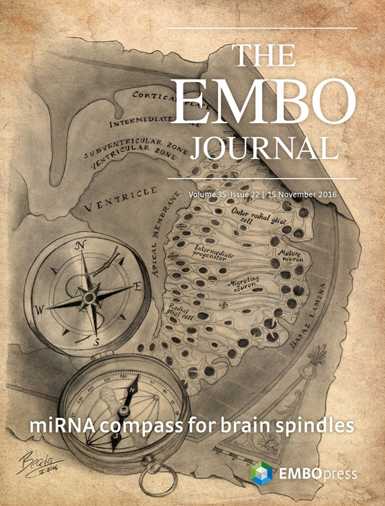
The angle of the mitotic spindle during cell division affects the decision-making of differentiation and thus ultimate position within the brain. This illustration was recently selected as the cover for an issue of EMBO Journal, accompanying a paper I was involved in during my Master’s thesis.
Research
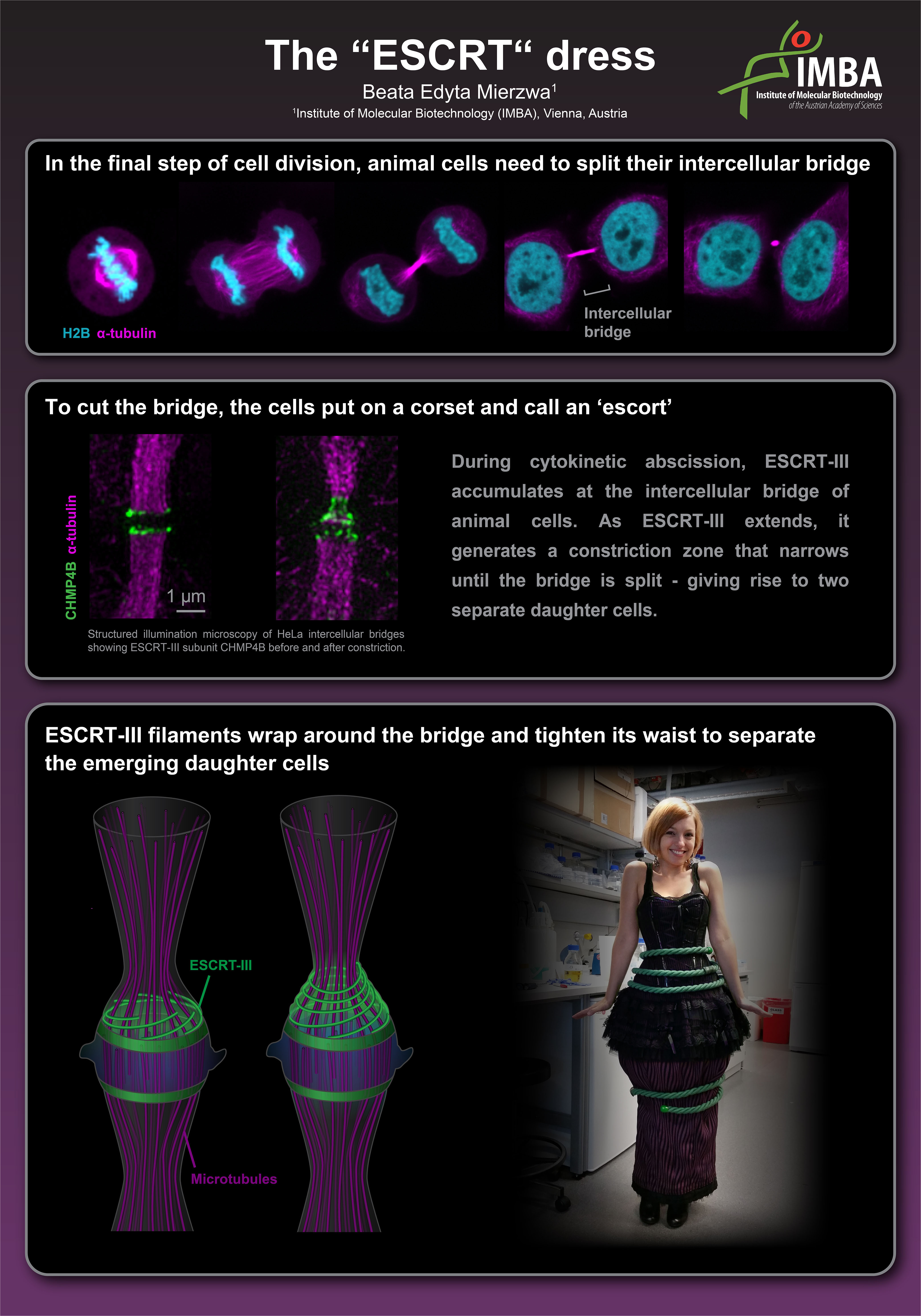
I work on cytokinetic abscission, the final step in cell division that separates the emerging daughter cells. Abscission is mediated by a machinery composed of the Endosomal Sorting Complex Required for Transport (ESCRT)-III. ESCRT-III is thought to form polymers that constrict the intercellular bridge until the membranes split. ESCRT‐III mediates membrane deformation and fission not only during cytokinesis, but also in various fundamental cellular processes, including multivesicular body formation, virus budding, plasma membrane repair, and nuclear envelope sealing. Current models propose that persistent filaments drive membrane constriction. However, whether ESCRT-III polymers exchange their subunits with soluble cytoplasmic pools—like other force-generating filament systems such as actin and tubulin—is not known.
My research project focuses on investigating the dynamics of this fascinating machinery, with the aim to shed light on how ESCRT-III polymers reorganize to drive membrane constriction and fission in a large diversity of cellular processes.
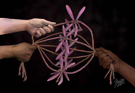
To illustrate how ESCRT-III splits the intercellular bridge during abscission, I created an “ESCRT” dress. This was a contribution to the Art & Science contest for the institutes at the Vienna Biocenter.
Check out my website (www.beatascienceart.com) for recent updates and a complete gallery.
All images © 2016 Beata Edyta Mierzwa
Guest posts are contributed by members of our community. The views expressed in guest posts are those of the author(s) and are not necessarily endorsed by the Genetics Society of America. If you'd like to write a guest post, e-mail communications@genetics-gsa.org.
View all posts by Guest Author »Read more in
-
In Memoriam: Ellsworth Herman Grell (1932–2023), a pioneer of Drosophila genome engineering and annotation
Ellsworth (Ed) Grell blessed the Drosophila community through three enduring legacies: as a pioneer of chromosome mechanics, as a primary organizer and synthesizer of genetic knowledge in Drosophila, and as a graceful mentor to those fortunate to have known him personally. Ed grew up in rural Nebraska, completed his undergraduate studies at Iowa State, and…
-

Congratulations to the #Fungal24 Poster Award winners!
We are pleased to announce the recipients of the GSA Poster Awards for posters presented at the 32nd Fungal Genetics Conference! Undergraduate and graduate student members of GSA were eligible for the awards, and a hard-working team of judges made the determinations. Congratulations to all! Felicia Ebot Ojong, The University of Georgia My research is focused…
-
Poster presentation tips for TAGC 2024
You’ve been selected to present a poster at The Allied Genetics Conference 2024 in March—you’ve celebrated, made plans to attend, now what? This is an exciting opportunity to showcase your research and engage with fellow members of the genetics community, so you want to make sure you’re prepared. We wanted to offer you some tips…
-

Maximize your TAGC 2024 experience
A guide to all that National Harbor & DC have to offer Are you joining us for The Allied Genetics Conference 2024 in March? Make the most of your #TAGC24 experience in National Harbor! We know the science will keep you busy, but you deserve to unwind and have some fun, so we’ve curated a…
-
Early Career Leadership Spotlight: Sarah Petrosky
We’re taking time to get to know the members of the GSA’s Early Career Scientist Committees. Join us to learn more about our early career scientist advocates. Sarah PetroskyMultimedia SubcommitteeUniversity of Pittsburgh Research Interest I am interested in understanding adaptation that has been happening recently in populations by dissecting the ways that genes underlying an adaptation…
-
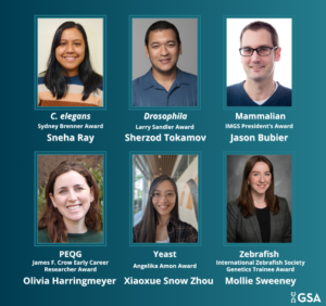
TAGC 2024 Early Career Award Winners
GSA is pleased to announce the winners of the early career awards presented at The Allied Genetics Conference 2024. These awards are specific to particular TAGC communities and recognize early career scientists’ outstanding work on their respective research organisms. The awardees will present their talks in keynote sessions at TAGC 2024. Don’t miss the opportunity…
-

Preeminent geneticists recognized with revamped GSA Awards
In 2022, GSA’s Board of Directors launched an audit to review the five major awards conferred by the Society. Today, we are thrilled to announce the recipients of the reimagined GSA Awards, including the new Genetics Society of America Early Career Medal. The scientists honored this year are recognized by their peers for their outstanding…
-
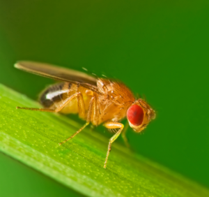
Fly Board funds outreach programs to spread the word about Drosophila research
In 2020, the Fly Board voted to use part of its reserve fund to support efforts to increase trainee participation as well as equity and diversity in the Drosophila community. An awards committee decides how the money will be spent each year, and from 2020–2022, the committee posted a very broad call for applications from…
-

New members of the GSA Board of Directors: 2024–2026
We are pleased to announce the election of four new leaders to the GSA Board of Directors: 2024 Vice President/2025 President Brenda Andrews Professor, University of Toronto It’s an honor to continue my association with the Society by serving as Vice President of the Board of Directors. I have broad knowledge of the ongoing activities…
-
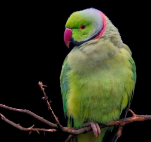
Parrot plumage study aids breeders and endangered natural populations
Yellow coloration is maladaptive in the wild but prized by breeders. People have long been fascinated with birds, which exhibit one of the widest ranges of coloration among vertebrates. Parrots, in particular, have captivated humans by their ability to mimic human speech and spectacular plumage. Brightly colored feathers are used primarily to attract mates, intimidate…
-
Early Career Leadership Spotlight: Sarah Gilmour
We’re taking time to get to know the members of the GSA’s Early Career Scientist Committees. Join us to learn more about our early career scientist advocates. Sarah GilmourMultimedia SubcommitteeStowers Institute for Medical Research Research Interest Questions of evolution have always fascinated me. I am extremely fortunate to be starting out my research career in a…